Brain anatomy is the foundation for understanding how this remarkable organ controls every function in your body. From breathing and balance to memory, emotion, and complex decision-making, each region of the brain plays a highly specialized role that keeps you alive and functioning every day.
Brain Anatomy: Understanding the Major Structures and Their Roles
For Canadians looking to learn more about their health, knowing the major parts of the brain can help you better understand neurological conditions, communicate with your doctor, and make informed decisions about your care. This guide walks you through the cerebral cortex lobes, brain stem, limbic system, and other key brain structures and functions so you can see exactly how each area contributes to your well-being.
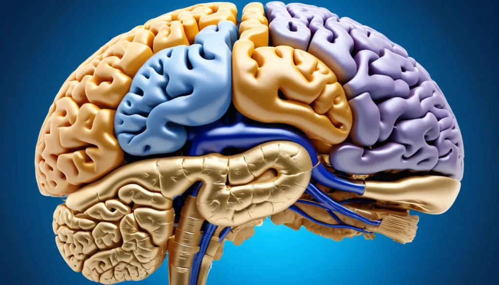
Brain Anatomy: The Cerebral Cortex and Its Lobes
Major Brain Anatomy Structures and Their Functions
| Brain Structure | Location | Primary Functions | Associated Conditions if Damaged |
|---|---|---|---|
| Frontal Lobe | Front of the brain, behind the forehead | Decision-making, personality, voluntary movement, speech production (Broca’s area) | Personality changes, impaired judgment, speech difficulties, paralysis |
| Temporal Lobe | Sides of the brain, near the temples | Language comprehension, memory formation, hearing, emotional responses | Memory loss, language disorders (Wernicke’s aphasia), hearing impairment |
| Parietal Lobe | Upper middle area of the brain | Sensory processing, spatial awareness, reading, and mathematical reasoning | Loss of sensation, difficulty with coordination, spatial disorientation |
| Occipital Lobe | Back of the brain | Visual processing, colour recognition, depth perception, object identification | Visual field loss, cortical blindness, difficulty recognizing objects |
| Cerebellum | Base of the brain, below the cerebrum | Balance, coordination, fine motor control, posture regulation | Ataxia, tremors, loss of balance, impaired coordination |
| Brain Stem | Lower portion connecting brain to spinal cord | Controls breathing, heart rate, blood pressure, sleep-wake cycles | Coma, locked-in syndrome, disrupted autonomic functions, death if severely injured |
The cerebral cortex is the outer layer of the brain. It is only 1.5 to 5 mm thick, but it handles some of the most complex tasks in the body. It receives and processes sensory information and is divided into four main lobes.
Each lobe has a distinct role. Together, they shape your personality, intelligence, and ability to interact with the world around you. According to Mayo Clinic, damage to specific lobes can cause very different health problems depending on location.
The Frontal Lobes
The frontal lobes sit at the front of the cerebral cortex. They handle decision-making, planning, reasoning, and impulse control. They also manage motor functions and short-term memory.
In addition, the frontal lobes play a key role in your personality. Injuries to this area can change how a person behaves and makes choices.
The Parietal Lobes
The parietal lobes sit behind the frontal lobes, above the occipital lobes. They process sensory information such as touch, pain, and spatial awareness. They also support speech and visual perception.
The Temporal Lobes
The temporal lobes are located on the sides of the brain, below the lateral sulcus. They handle hearing, memory, speech, and emotional responses. Damage here can affect a person’s ability to recognise faces or understand language.
The Occipital Lobes
The occipital lobes are at the back of the brain. Their main job is to process visual information, including colour recognition. Therefore, any injury to this area can cause problems with sight.
Brain Anatomy: The Cerebellum and Brain Stem
Two of the most vital structures in brain anatomy are the cerebellum and the brain stem. Both sit near the base of the skull and control functions your body performs automatically.
The Cerebellum
The cerebellum sits above the brain stem, just below the occipital lobes. It coordinates fine motor movements and helps you maintain balance. It also regulates muscle tone.
For example, when you reach for a glass of water, the cerebellum ensures your hand moves smoothly and accurately. Diseases that affect the cerebellum can cause unsteady movements and poor coordination.
The Brain Stem
The brain stem connects the brain to the spinal cord. It is made up of three parts: the midbrain, the pons, and the medulla oblongata. This structure controls many automatic functions that keep you alive.
These functions include breathing, heart rate, blood pressure, digestion, and alertness. The brain stem also contains most of the cranial nerves. As a result, damage here can affect many systems at once.
The Basal Ganglia and Cerebral Hemispheres
The basal ganglia are a group of structures located deep within the cerebral hemispheres. They are involved in voluntary movement and thinking. They are made up of the striatum, the subthalamic nuclei, and the substantia nigra.
However, the basal ganglia are perhaps best known because of the diseases linked to them. Parkinson’s disease and Huntington’s disease both involve damage to these structures. Health Canada recognises these as serious neurological conditions that affect many Canadians.
The cerebral hemispheres make up the largest part of the brain. They are divided into the left and right sides, connected by a thick band of fibres called the corpus callosum. The corpus callosum allows the two sides to share information quickly and efficiently.
The Limbic System: Emotion, Memory, and Behaviour
The limbic system is a group of structures deep inside the brain. It plays a central role in emotion, memory, and behaviour. Understanding this part of brain anatomy helps explain why stress and trauma can have lasting effects.
The Amygdala
The amygdala processes emotional responses, including fear and pleasure. It also plays a role in hormone secretion and memory formation. When you feel anxious or startled, the amygdala is likely involved.
The Hippocampus
The hippocampus sends information to the cerebral hemispheres for long-term storage. It also retrieves memories when needed. Furthermore, it is one of the first areas affected by Alzheimer’s disease, which can cause significant memory loss.
The Hypothalamus
The hypothalamus controls many important body functions. These include body temperature, hunger, thirst, and hormonal balance. It acts like a command centre that keeps your internal environment stable.
Other Limbic Structures
The cingulate gyrus is a fold of brain tissue involved in sensory impulses related to emotion. It also helps regulate aggressive behaviour. The fornix is an arched band of nerve fibres that connects the hippocampus to the hypothalamus.
Broca’s Area and Key Sulci
Broca’s area is found in the left frontal lobe, around the opercular and triangular regions of the inferior frontal gyrus. It controls the facial neurons involved in speech production and plays a role in understanding language. Damage to Broca’s area can cause expressive aphasia, meaning a person struggles to speak clearly.
Two important sulci, or grooves, also shape brain anatomy. The central sulcus of Rolando separates the frontal and parietal lobes. The lateral sulcus of Sylvius separates the parietal and temporal lobes. These deep grooves increase the brain’s surface area significantly.
The 12 Cranial Nerves
There are 12 pairs of cranial nerves that originate in the brain. They exit through the skull and serve the head, neck, and trunk. Each pair has a specific function.
These functions include directing sensory impulses, controlling eye movements, managing facial sensations, supporting hearing and speech, and regulating swallowing and breathing. According to Healthline, problems with cranial nerves can signal serious neurological conditions.
Optic, oculomotor, and trochlear nerves originate at the front of the brain and control vision and eye movement.
Trigeminal, abducens, and facial nerves start at the pons and manage facial sensation and movement.
Vestibulocochlear nerve originates at the inner ear and connects to the pons, supporting hearing and balance.
Glossopharyngeal, vagus, accessory, and hypoglossal nerves attach to the medulla oblongata and control swallowing, speech, and other functions.
When to See a Doctor
Many brain conditions develop slowly. You may not notice symptoms right away. However, some warning signs need prompt medical attention.
See your family doctor or visit a walk-in clinic if you experience sudden severe headaches, confusion, memory loss, difficulty speaking, vision changes, or loss of balance. These could be signs of a neurological problem that needs evaluation. Your family doctor can refer you to a neurologist through your provincial health plan if needed.
If symptoms appear suddenly and severely, call 911 or go to the nearest emergency department immediately. Do not wait. Early treatment can make a major difference in outcomes for stroke, brain injury, and other serious conditions.
This article is for general information only. Always speak with a qualified healthcare provider for personal medical advice and diagnosis.
What are the main structures in brain anatomy?
The main structures in brain anatomy include the cerebral cortex, cerebellum, brain stem, basal ganglia, limbic system, and corpus callosum. Each structure controls different functions, from movement and balance to memory and emotion. Together, they work as a highly coordinated system.
What does the cerebral cortex do?
The cerebral cortex is the outer layer of the brain and plays a key role in brain anatomy. It processes sensory information, controls motor functions, and supports intelligence, personality, and planning. It is divided into four lobes: frontal, parietal, temporal, and occipital.
What part of the brain controls memory?
The hippocampus, part of the limbic system, is the primary structure involved in forming and retrieving memories. The frontal lobes also contribute to short-term memory and decision-making. Damage to these areas, as seen in Alzheimer’s disease, can cause significant memory problems.
What diseases are linked to the basal ganglia?
The basal ganglia are closely linked to Parkinson’s disease and Huntington’s disease. Both conditions involve damage to these deep brain structures, affecting voluntary movement and thinking. If you have concerns about movement disorders, speak with your family doctor or visit a walk-in clinic.
How many cranial nerves does the human brain have?
The human brain has 12 pairs of cranial nerves, a key part of understanding brain anatomy. They originate in the brain and control functions like vision, hearing, facial movement, swallowing, and breathing. Problems with cranial nerves may require assessment by a neurologist through your provincial health plan.
What is the function of the brain stem?
According to Mayo Clinic’s overview of brain structures, this information is supported by current medical research.
For more information, read our guide on understanding your white blood cell count results in Canada.
The brain stem connects the brain to the spinal cord and controls automatic functions essential for survival. These include breathing, heart rate, blood pressure, and digestion. It is made up of the midbrain, pons, and medulla oblongata.
Key Takeaways
Brain anatomy includes many specialised structures, each with unique and critical functions.
The cerebral cortex, divided into four lobes, handles thinking, sensation, vision, hearing, and movement.
The cerebellum controls coordination and balance, while the brain stem manages automatic survival functions.
The basal ganglia are involved in voluntary movement and are linked to Parkinson’s and Huntington’s diseases.
The limbic system, including the hippocampus and amygdala, governs memory, emotion, and behaviour.
There are 12 pairs of cranial nerves that control many functions in the head, neck, and trunk.
If you notice sudden neurological symptoms, see your family doctor, visit a walk-in clinic, or call 911 in an emergency.
Frequently Asked Questions
What is brain anatomy and why is it important?
Brain anatomy refers to the physical structure of the brain, including its regions, lobes, and neural pathways. Understanding brain anatomy is essential for diagnosing neurological conditions, guiding surgical procedures, and interpreting symptoms. It helps doctors identify which brain area is affected following injury, stroke, or disease in Canadian patients.
What are the key structures of brain anatomy and their functions?
The brain’s key structures include the cerebrum (thinking and movement), cerebellum (balance and coordination), brainstem (breathing and heart rate), hippocampus (memory), and amygdala (emotions). Each region plays a distinct role, and damage to any area can cause specific neurological symptoms depending on the location and severity of injury.
What are the symptoms of a brain injury or neurological problem?
Common symptoms include sudden severe headache, confusion, memory loss, vision changes, slurred speech, weakness on one side of the body, seizures, and loss of coordination. These symptoms may indicate stroke, traumatic brain injury, or other serious neurological conditions requiring immediate medical attention at a Canadian emergency department.
Can brain damage be treated or reversed in Canada?
Some brain damage can be treated and partially reversed depending on cause and severity. Stroke treatment is most effective within hours of onset. Rehabilitation including physiotherapy, occupational therapy, and speech therapy helps many Canadians recover lost function. Early diagnosis and access to specialized neurological care significantly improve long-term recovery outcomes.
When should I see a doctor about brain or neurological symptoms?
See a doctor immediately if you experience sudden severe headache, confusion, one-sided weakness, vision loss, slurred speech, or loss of consciousness. These are potential stroke or brain injury warning signs. In Canada, call 911 or visit your nearest emergency department without delay, as early treatment dramatically improves outcomes.