
Craniopharyngioma: Symptoms, Diagnosis & Treatment Canada
Share
Craniopharyngioma is a rare, slow-growing brain tumour that develops near the pituitary gland at the base of the brain. Although classified as benign, this tumour can cause serious complications over time, including vision loss, hormonal imbalances, and cognitive difficulties. It affects both children and adults, making early detection essential for better outcomes.
What Is a Craniopharyngioma and How Does It Affect the Brain?
Understanding craniopharyngioma symptoms is the first step toward timely diagnosis and effective care. For Canadians, knowing the signs, diagnostic procedures, and treatment options available through provincial health systems can make a significant difference. This guide covers everything you need to know about craniopharyngioma, from how it is diagnosed with imaging and blood tests to surgical and radiation-based treatment options offered across Canada.
What Is a Craniopharyngioma?
| Treatment Option | Description | Benefits | Key Considerations |
|---|---|---|---|
| Surgical Resection | Removal of the craniopharyngioma tumour through open craniotomy or minimally invasive endoscopic approach via the nasal passage | Potential for complete tumour removal; immediate relief of pressure on surrounding structures | Risk of damage to pituitary gland, optic nerves, and hypothalamus; may require hormone replacement therapy post-surgery |
| Radiation Therapy | Targeted high-energy beams used to destroy remaining tumour cells following surgery or as a primary treatment when surgery is not feasible | Effective for residual or recurrent tumours; stereotactic radiosurgery (e.g., Gamma Knife) offers precision with minimal damage to surrounding tissue | Risk of long-term cognitive effects, particularly in children; not suitable for tumours adjacent to critical structures in some cases |
| Intracavitary Therapy | Direct injection of chemotherapy agents (e.g., bleomycin or interferon-alpha) into cystic portions of the tumour | Targets cystic craniopharyngioma directly; may reduce tumour size before surgery | Limited to cystic tumour types; risk of chemical leakage causing damage to adjacent brain tissue |
| Hormone Replacement Therapy | Long-term management of endocrine deficiencies caused by craniopharyngioma or its treatment, including growth hormone, cortisol, and thyroid hormone supplementation | Restores hormonal balance; improves quality of life and supports normal development in paediatric patients | Requires lifelong monitoring and adjustment; available through endocrinology services across Canada’s provincial health systems |
| Watchful Waiting | Active surveillance with regular MRI imaging for small, asymptomatic craniopharyngiomas that are not immediately threatening critical structures | Avoids treatment-related side effects; appropriate for stable, slow-growing tumours in select patients | Requires consistent follow-up imaging every 6–12 months; intervention may still be needed if tumour progresses |
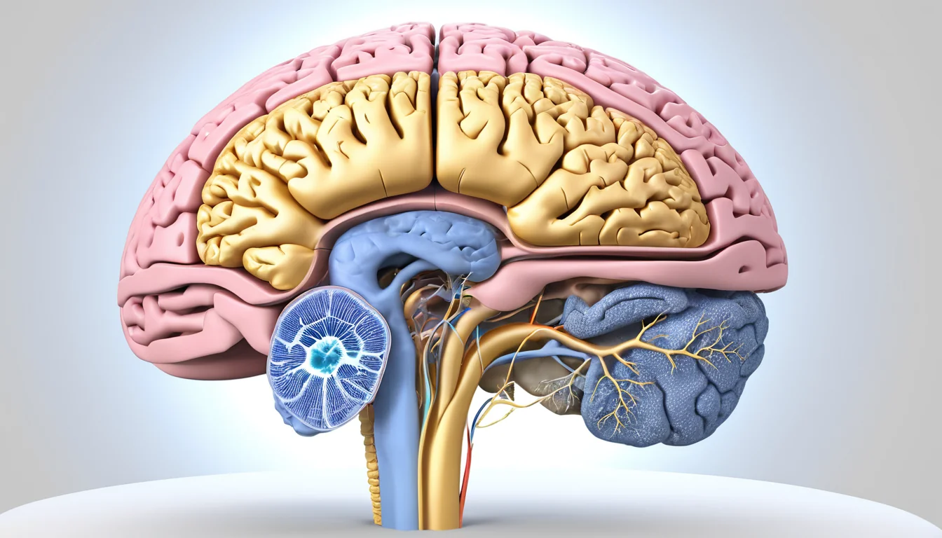
A craniopharyngioma is a non-cancerous (benign) tumour that develops near the pituitary gland. The pituitary gland sits in a small bony hollow at the base of the skull, called the sella turcica, or “Turkish saddle.” Doctors believe these tumours grow from leftover embryonic tissue that was important in forming the pituitary gland during fetal development.
These tumours grow slowly but can cause local damage as they expand. They often contain areas of calcification (hardened calcium deposits) and fluid-filled cysts. Their texture can range from mostly solid to mostly cystic. Craniopharyngioma can affect both children and adults, including older Canadians.
According to the Mayo Clinic’s overview of craniopharyngioma, this tumour accounts for a small but significant portion of all brain tumours diagnosed each year.
Where Does a Craniopharyngioma Develop?
A craniopharyngioma most often forms in and around the pituitary gland. It usually extends upward into the hypothalamus, a critical area of the brain that controls many body functions. The optic nerves — the nerves that carry visual signals from your eyes to your brain — also run through this region.
As the tumour grows, it can press on nearby structures. This pressure causes many of the symptoms people experience. The tumour can extend in several directions:
- Forward (anterior): Into the prechiasmatic and subfrontal spaces near the front of the brain.
- Backward (posterior): Toward the prepontine area, the third ventricle, and even the posterior fossa.
- Sideways (lateral): Into the subtemporal spaces on either side of the brain.
The location of the tumour plays a large role in which symptoms a person develops. Therefore, no two cases of craniopharyngioma look exactly alike.
Symptoms of Craniopharyngioma
Because craniopharyngiomas grow slowly, symptoms often develop gradually over months or even years. Many people do not notice anything unusual at first. However, as the tumour grows, symptoms become more noticeable.
General Symptoms
Headaches are one of the most common general symptoms. People may also feel fatigued, confused, or generally unwell. These symptoms are easy to overlook or mistake for other conditions.
Vision Problems
The tumour’s closeness to the optic nerves means that vision changes are very common. People may notice blurry vision, blind spots, or loss of peripheral (side) vision. In more serious cases, significant vision loss can occur. For this reason, an eye examination is often an important part of the diagnostic process.
Hormonal Symptoms
The pituitary gland and the hypothalamus control many important hormones. When a craniopharyngioma damages these structures, hormone deficiencies can develop. These hormonal problems can affect many systems in the body.
Common hormone-related symptoms include:
- Growth hormone deficiency: Children may experience slowed growth. Adults may notice changes in body composition.
- Adrenal insufficiency: This can cause low blood pressure, low blood sugar (hypoglycaemia), extreme tiredness, and confusion.
- Hypothyroidism: Symptoms include weight gain, sensitivity to cold, and fatigue.
- Diabetes insipidus: This condition causes extreme thirst and excessive urination due to a lack of a hormone that regulates fluid balance.
- Reproductive hormone problems: Men may experience erectile dysfunction. Women may have irregular or absent menstrual periods. Younger patients may experience delayed puberty.
According to Healthline’s guide on craniopharyngioma, hormone problems affect the majority of people diagnosed with this tumour, making hormonal testing a key part of diagnosis and follow-up care.
Behavioural and Cognitive Symptoms
Larger tumours can affect the hypothalamus and surrounding brain tissue more broadly. As a result, some people experience changes in behaviour, memory, or mood. These effects can significantly impact daily life and quality of life.
How Is Craniopharyngioma Diagnosed?
Diagnosing a craniopharyngioma involves several steps. Your doctor will start with a thorough neurological examination to assess your reflexes, vision, coordination, and mental function.
Imaging Tests
A CT scan or MRI (magnetic resonance imaging) of the brain is usually the next step. These scans show the size, shape, and location of the tumour clearly. A craniopharyngioma has a fairly distinctive appearance on imaging, with its mix of solid tissue, cysts, and calcium deposits. However, doctors must also rule out other tumours that can appear in the same area, such as a pituitary adenoma or a meningioma.
Tissue Biopsy
A precise diagnosis requires a tissue sample. A specialist called a pathologist examines the tissue under a microscope. This confirms whether the tumour is truly a craniopharyngioma and rules out other types of growths.
Hormone Blood Tests
Almost all patients with a tumour in this region need hormone blood tests. These tests check the levels of several pituitary hormones and related hormones. The results help identify any hormone deficiencies caused by the tumour pressing on the pituitary gland or hypothalamus. Early identification of hormone problems allows doctors to start hormone replacement therapy promptly.
Eye Examination
A detailed eye exam is usually recommended. This helps determine whether the tumour has affected the optic nerves and how much vision may be impaired. An ophthalmologist (eye specialist) typically performs this assessment.
For more information on how brain tumours are diagnosed in Canada, visit Health Canada’s official health information portal.
Treatment Options for Craniopharyngioma
Treatment for craniopharyngioma is complex and usually involves a team of specialists. The goal is to remove or reduce the tumour while protecting surrounding brain structures as much as possible.
Surgery
Surgery is typically the first-line treatment. A neurosurgeon works to remove as much of the tumour as safely possible. Because the tumour sits close to vital brain structures, complete removal is not always achievable without causing harm. In these cases, surgeons aim for a safe partial removal.
Radiation Therapy
Radiation therapy is often used after surgery. It targets any remaining tumour tissue to prevent regrowth. Modern techniques, such as stereotactic radiosurgery (for example, Gamma Knife), deliver precise doses of radiation with minimal damage to surrounding tissue.
Hormone Replacement Therapy
Many patients need long-term hormone replacement after treatment. This is because surgery and radiation can further damage the pituitary gland or hypothalamus. Hormone replacement helps manage deficiencies in thyroid hormone, cortisol, growth hormone, and others. An endocrinologist (hormone specialist) typically manages this ongoing care.
Cyst Drainage
In some cases, doctors can drain the fluid-filled cysts directly. This is sometimes done using a small catheter (tube) placed into the cyst. It can relieve pressure quickly, though it may not be a permanent solution.
When to See a Doctor
If you or a family member notices persistent headaches, unexplained vision changes, extreme fatigue, or unusual hormonal symptoms, do not wait. These symptoms deserve prompt medical attention. Start by calling your family doctor or visiting a walk-in clinic. They can assess your symptoms, order initial tests, and refer you to a specialist if needed.
Most provincial health plans in Canada cover the diagnostic tests and specialist referrals needed to investigate these kinds of symptoms. You do not need to navigate this alone. Your family doctor or walk-in clinic is the best first point of contact.
Please speak with a qualified healthcare provider before drawing any conclusions about your health. Only a doctor can properly evaluate your symptoms and recommend the right course of action.
Frequently Asked Questions About Craniopharyngioma
Is craniopharyngioma cancerous?
No, a craniopharyngioma is generally considered a benign (non-cancerous) tumour. However, it can still cause serious harm because of its location near vital brain structures. Even though it does not spread to other parts of the body like a malignant tumour, a craniopharyngioma requires prompt medical treatment.
Who is most likely to develop a craniopharyngioma?
Craniopharyngioma can develop at any age, but it is most commonly diagnosed in children between 5 and 14 years old and in adults between 50 and 74 years old. It affects males and females roughly equally. There are no known lifestyle or environmental risk factors clearly linked to craniopharyngioma development.
Can craniopharyngioma come back after treatment?
Yes, craniopharyngioma can recur (come back) after treatment, especially if the entire tumour could not be safely removed during surgery. This is why regular follow-up MRI scans and hormone checks are so important after treatment. Your medical team will set up a long-term monitoring plan to detect any regrowth early.
What are the long-term effects of craniopharyngioma?
Long-term effects of craniopharyngioma can include hormone deficiencies, vision problems, changes in weight, memory difficulties, and changes in behaviour. Many of these effects result from both the tumour itself and from the treatments used to remove it. Long-term hormone replacement therapy and regular specialist follow-up help manage these effects effectively.
How is craniopharyngioma different from a pituitary adenoma?
Both craniopharyngioma and pituitary adenoma are tumours near the pituitary gland, but they are different types. A pituitary adenoma grows directly from the pituitary gland’s own cells, while a craniopharyngioma grows from embryonic tissue remnants near the gland. They also look different on brain scans and require somewhat different treatment approaches. A tissue biopsy is the most reliable way to tell them apart.
Is craniopharyngioma covered under provincial health plans in Canada?
According to Mayo Clinic’s overview of craniopharyngioma, this information is supported by current medical research.
For more information, read our guide on pituitary tumours and their symptoms in Canada.
Yes, diagnosis and treatment of craniopharyngioma — including MRI scans, specialist consultations, surgery, and radiation therapy — are generally covered under provincial and territorial health plans across Canada. However, coverage details can vary by province. It is always a good idea to speak with your family doctor or provincial health authority to understand exactly what is covered in your region.
Key Takeaways
- A craniopharyngioma is a rare, slow-growing benign tumour near the pituitary gland at the base of the brain.
- It can affect your vision, hormones, and brain function as it grows and presses on surrounding structures.
- Symptoms develop slowly and may include headaches, vision changes, hormone deficiencies, and behavioural changes.
- Diagnosis involves MRI or CT scans, hormone blood tests, eye examinations, and a tissue biopsy.
- Treatment usually combines surgery, radiation therapy, and long-term hormone replacement therapy.
- Most provincial health plans in Canada cover the necessary tests and treatments.
- If you notice concerning symptoms, contact your family doctor or visit a walk-in clinic promptly.
- Always consult a qualified healthcare provider for personal medical advice.
Frequently Asked Questions
What is a craniopharyngioma?
A craniopharyngioma is a rare, benign (non-cancerous) brain tumour that develops near the pituitary gland at the base of the skull. It grows from remnant embryonic tissue and most commonly affects children aged 5–14 and adults over 50. Despite being benign, it can cause serious complications by pressing on surrounding brain structures.
What are the symptoms of craniopharyngioma?
Common symptoms include persistent headaches, vision problems, unexplained weight gain, stunted growth in children, excessive thirst and urination, fatigue, and hormonal imbalances. As the tumour grows, it can increase intracranial pressure, causing nausea and vomiting. Symptoms develop gradually, often making early diagnosis challenging.
How is craniopharyngioma treated in Canada?
Treatment typically involves surgical removal of the tumour, often followed by radiation therapy to eliminate remaining cells. Canadian neurosurgeons may use minimally invasive endoscopic techniques. Hormone replacement therapy is frequently required post-treatment due to pituitary gland damage. Treatment plans are individualized and managed by multidisciplinary teams at major Canadian cancer centres.
Can craniopharyngioma be prevented?
No, craniopharyngioma cannot be prevented. It develops from embryonic tissue remnants during fetal development, meaning risk factors are not related to lifestyle or environmental choices. There are no known modifiable risk factors. Early detection through prompt medical evaluation of symptoms offers the best opportunity for successful treatment outcomes.
When should you see a doctor for craniopharyngioma symptoms?
See a doctor promptly if you or your child experience persistent headaches, sudden vision changes, unexplained weight gain, abnormal thirst, or delayed growth and puberty. These symptoms can indicate a brain tumour or hormonal disorder requiring urgent evaluation. Early diagnosis significantly improves treatment success and quality of life outcomes.
About the Author
Dr. Sarah Mitchell, MDDr. Sarah Mitchell is a board-certified family physician with over 15 years of clinical experience. She completed her MD at the University of Toronto and her residency at Toronto General Hospital. Dr. Mitchell specializes in preventive medicine and chronic disease management. She is a member of the College of Family Physicians of Canada (CFPC) and has published over 30 peer-reviewed articles on preventive health care.
View all articles →